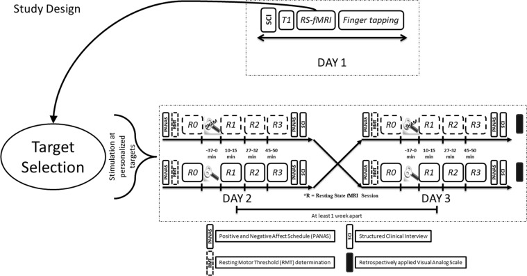
Figure 8.
Study design – On day 1, after a Structured Clinical Interview (SCI) and obtaining informed consent, we acquired a T1 weighted structural image, rsfMRI and finger tapping task based fMRI. During rsfMRI, we instructed subjects to look at a fixation cross presented on a black background. The rsfMRI data was used for personalized target selection and the personalized target was then identified in T1 structural for stimulation using online neuronavigation. rsfMRI sessions were followed by an index finger tapping session (intermittent finger tapping when presented with a green or a red dot), data from which was used to determine the motor cortex location used for setting the resting motor threshold (RMT). At the beginning of experiment on day 2 and day 3, we asked each subject to complete the Positive and Negative Affect Schedule (PANAS). This was followed by resting motor threshold determination. We obtained a baseline rsfMRI scan (R0), and then delivered HF rTMS stimulation (either real or sham in a pseudo-randomized and counterbalanced way) to the subject at the pre-selected target, guided by online neuronavigation. Post HF rTMS three additional rsfMRI scans (R1, R2, and R3) were obtained over a course of 50 minutes: at 10 minutes, 27 minutes and 45 minutes to detect effects on brain resting state functional connectivity. The subjects again completed the PANAS immediately after leaving the MR scanner. This allowed us to document any short term changes in the self-rated emotional state. The subjects completed a SCI again at the end of experiment on both day 2 and day 3. To assess the effectiveness of sham blinding, we also retrospectively collected information about the perceived effects of stimulation on scalp sensation and mental state using a visual analog scale (VAS).

