
Figure 1.
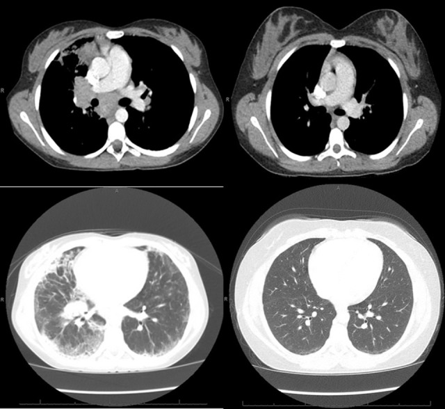
Left: CT of the chest obtained around the time of clinical presentation of GLILD, showing mediastinal and hilar lymphadenopathy, peripheral interlobular septal thickening, peripheral consolidation, and ground glass opacities, more prominent on the right lung than left. Right: CT of the chest obtained after treatment with 4 doses of rituximab and azathioprine showing resolution of the lymphadenopathy, consolidation and septal thickening and near resolution of the ground glass opacities.