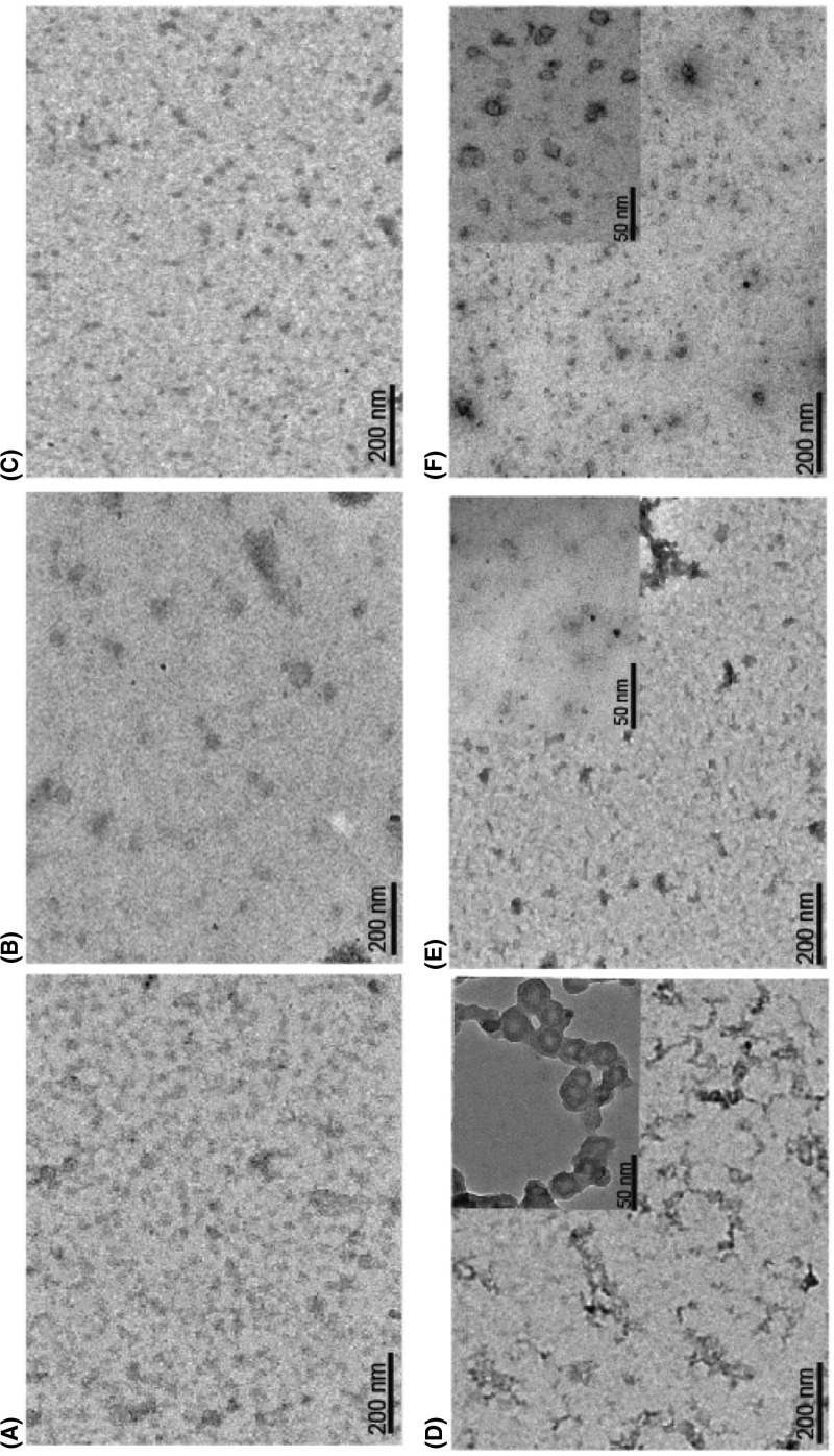
Figure 3. Electron micrographs showing reduction in the size and shape of AT polymers by sorbitol and trehalose.
AT Polymers were formed by heating 10 µM of the protein at 60°C for 90 min. Samples were stained negatively with 1.5% (w/v) uranyl acetate, and viewed with a magnification of up to ×50000. AT incubated at (A) 0 min, (D) 90 min; AT incubated in the presence of 1.5 M sorbitol at (B) 0 min and (E) 90 min and in the presence of 1 M trehalose at (C) 0 min and (F) 90 min. Insets in D–F shows enlarged view of polymers.

