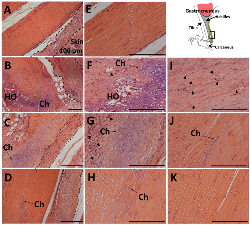
Figure 3.
Histological analysis of the tendon sections revealed heterotopic bone in SP- and chondrocyte formation in CGRP- or SP + CGRP-delivered tendons. Low magnification images of the Hematoxylin and Eosin (H&E) staining of mid-tendon region in (A) Control, (B) SP, (C) CGRP, (D) SP + CGRP. (E–H), (I–K). Higher magnification of the same H&E stained sections, zoomed into the tendon. Schematic shows the orientation of the images (tendon toward the skin- left to right). ADM is visible next to the tendon (marked with dashed lines), which was infiltrated by mononuclear cells. SP induced degenerative changes in the Achilles, including disorganized collagen, vascularization (marked by black arrowheads), and heterotopic ossification (HO) surrounded by chondrocytes (Ch). CGRP and SP + CGRP led to chondrocyte differentiation (Ch). CGRP also induces vascularization (Black arrowheads). Control tendons did not who show any pathological changes. Cellular proliferation/infiltration was observed around the tendon borders (black and white arrowheads). These regions potentially correspond to paratenon with vascular growth. The proliferation might be a result of surgical trauma. All scale bars measure 100 μm.

