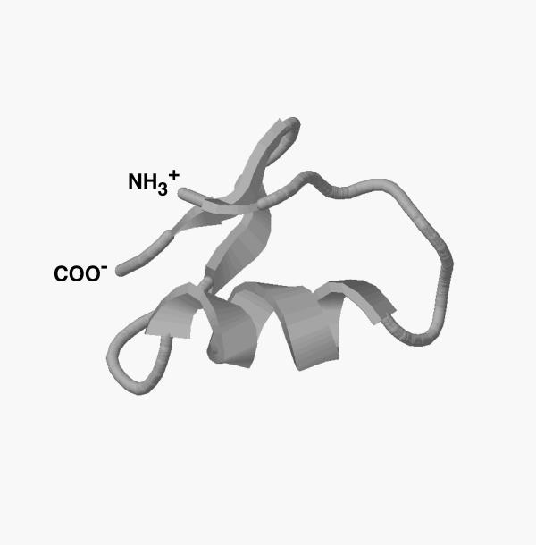
Figure 6.
The three-dimensional structure of Buta IT derived by Swiss-Model Protein Modeling Server using the NMR coordinates of chlorotoxin and insecotoxin I5A. The RasMol program is used to visualize the 3-D structure of the molecule. The carbon backbone trace of the molecule displays a basic scaffold composed of an α-helix and three β-strands similar to other short toxins.

