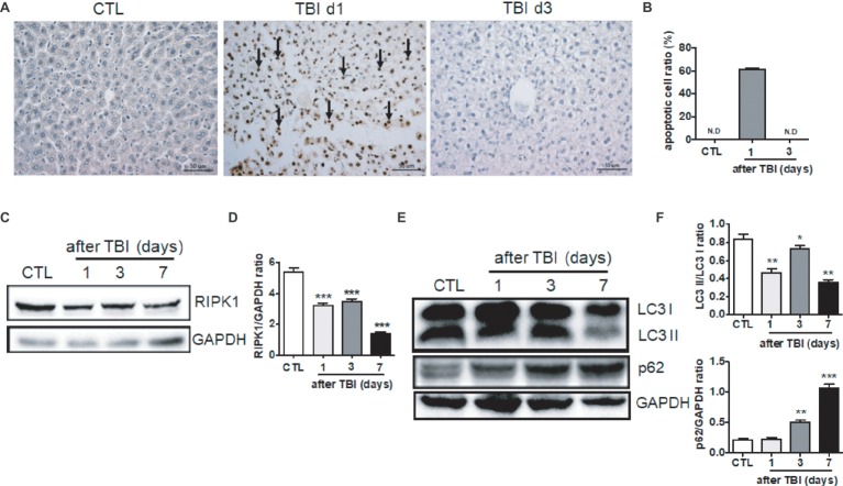
Figure 2.
Irradiation-induced cellular apoptosis in liver tissues. (A and B) Irradiated liver tissues were collected at days 1 and 3 after TBI. Representative pictures by TUNEL staining are shown (A, n = 6). Scale bar = 50 μm. Arrows indicate TUNEL-positive cells. The ratios of apoptotic cells in each field are presented in (B) (N.D: non-detected). (C and D) RIPK1 expression in liver tissues (n = 3). Expression of RIPK1 in liver tissues was detected by western blotting (C). GAPDH was used as a housekeeping control. Expression of RIPK1 was quantitated by ImageJ software (D). (E and F) LC3 I, LC3II, and p62 expression in liver tissues (n = 3). Expression of LC3 I, LC3II, and p62 (E) was detected by western blotting at days 1, 3, and 7 after irradiation. GAPDH was used as a housekeeping control. Expression of LC3 I, LC3II, and p62 (F) was quantitated by ImageJ software. The ratio of LC3 II/LC3 I was presented. *p < 0.05, **p < 0.01, ***p < 0.001 vs. CTL.

