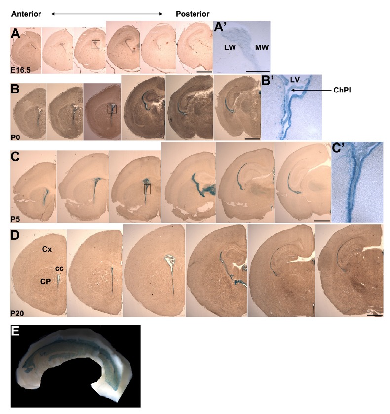
Fig. 1. Analysis of Anks1a expression during brain development.

(A–D) The whole brains of Anks1a heterozygous mice at the indicated embryonic or postnatal days were processed for cryostat sectioning. Coronal sections of the brain were subjected to X-gal staining to reveal LacZ-positive cells lining the lateral ventricle. cc, corpus callosum; ChPl, choroid plexus; CP, caudate putamen; Cx, cerebral cortex; LV, lateral ventricle; LW, lateral wall; MW, medial wall. Scale bars, 1 mm. The boxes in A–C were enlarged in A′–C′. Scale bar for A′, 250 μm. (E) The lateral wall (LW) of Anks1a heterozygous mice on P45 was subjected to whole mount X-gal staining. Note that ependymal cells in the LW display prominent X-gal staining. At least three embryos or mice were analyzed at each developmental stages and the results were reproducible.
