
Fig. 5. sh-PVT1 attenuated Ang II-induced PVT1 expression, ECM degradation, and inflammation in mice.
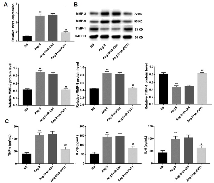
(A) Relative PVT1 expression in aortic tissues from mice four weeks after Ang II infusion in each group was examined by qRT-PCR. (B) The protein levels of MMP-2, MMP-9, and TIMP-1 in aortic tissues from mice four weeks after Ang II were examined by Western blot. GAPDH served as the loading control. (C) Four weeks after Ang II infusion, serum levels of the inflammatory cytokines TNF-α, IL-1β, and IL-6 were quantified using ELISA kits. n=15 for each group. ** P < 0.01 vs. NS group, and ##P < 0.01 vs. Ang II+sh-Ctrl group. NS: normal saline; Ang II: Angiotensin II; MMP-2: matrix metalloproteinase-2; MMP-9: matrix metalloproteinase-9; TIMP-1: tissue inhibitors of metalloproteinase-1; TNF-α: tumour necrosis factor-α; IL-1β: interleukin-1β; IL-6: interleukin-6.