
Fig. 1.
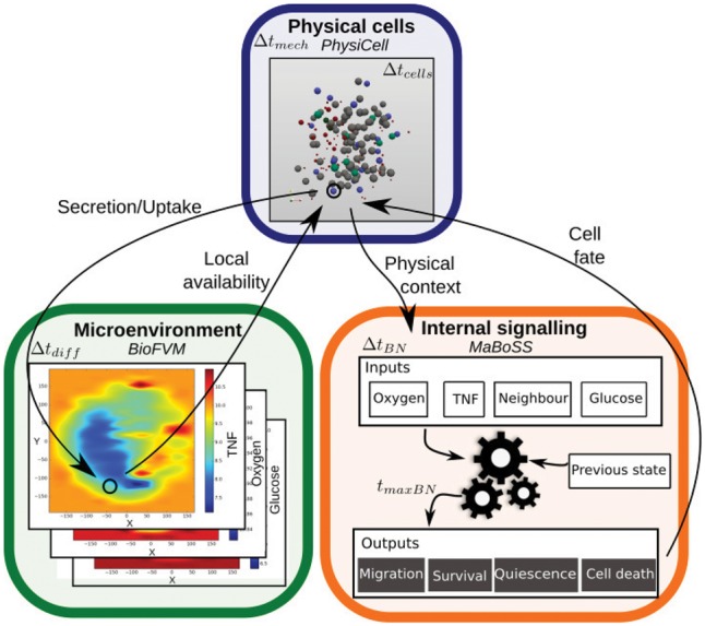
Schematic representation of PhysiBoSS. Three main parts are interconnected: the microenvironment representation in BioFVM (green, bottom left), allowing simulation of diffusing entities; the physical representation of cells as dynamic spheres in PhysiCell (blue, top); and the signalling modelling of each cell in MaBoSS (orange, bottom right) (Color version of this figure is available at Bioinformatics online.)