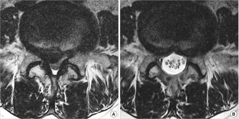
Fig. 7.

(A) Preoperative magnetic resonance imaging (MRI) showing the severe spinal stenosis combined with lumbar herniated nucleus pulposus. (B) Postoperative MRI taken 1 day after operation showing the sufficient decompression and adequate resection of lumbar herniated disk.
