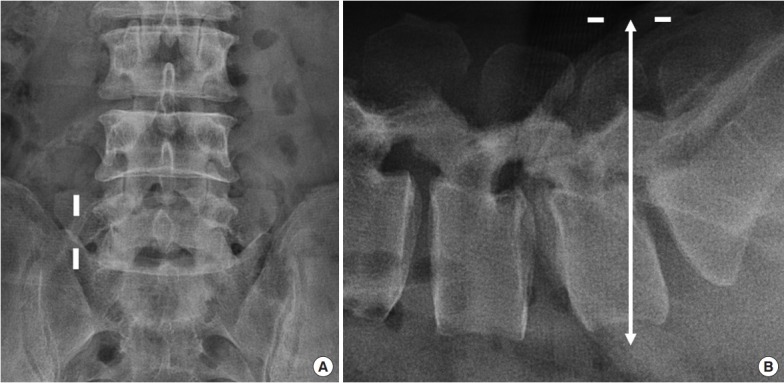
Fig. 3.

Skin incision points for the 2 portals. (A) Two channels were made at 1 cm lateral to the lateral border of the pedicle of L5–S1 in the anterior-posterior view. (B) Portals were made 1 cm below and 1 cm above the midpoint of the foramen (white arrow) in the lateral view. Generally, the upper portal was made around the pedicle of L5 and the lower portal was made around the upper endplate of S1 in the lateral C-arm view.
