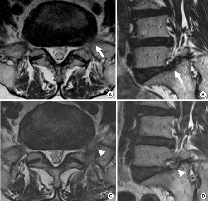
Fig. 4.

A 62-year-old female patient complained of left radicular leg pain. (A, B) The axial and oblique T2-weighted magnetic resonance images depict the extraforaminal entrapment of the L5 nerve root by disc protrusion and pseudo-arthrosis (white arrows). (C, D) After endoscopic surgery, the L5 nerve root was completely decompressed (white arrowheads) (Supplementary video clip 1).
