
Fig. 2.
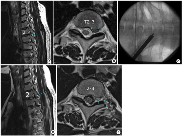
A 35-year-old female patient: (A) Preoperative sagittal view showing foraminal thoracic disc herniation (TDH) at the T2–3 level. (B) Preoperative axial view showing left foraminal TDH at the T3–4 level. (C) Intraoperative fluoroscopic view showing working channel is place at the foraminal area after foraminoplasty. (D) Postoperative sagittal view showing successful removal of disc herniation. (E) Postoperative axial view showing successful removal of disc herniation. Note that T2 nerve root is visible after decompression (arrow) and amount of foraminoplasty (arrowheads).