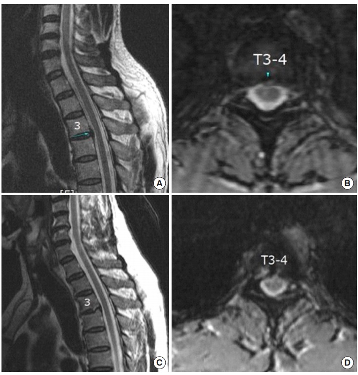
Fig. 3.

A 52-year-old male patient: (A) Preoperative sagittal view showing central thoracic disc herniation (TDH) at the T3–4 level. (B) Preoperative axial view showing central TDH at the T3–4 level. (C) Postoperative sagittal view showing successful removal of disc herniation. Note that the amount of foraminoplasty to access central part of disc. (D) Postoperative axial view showing successful removal of herniated disc.
