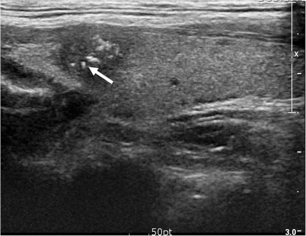
Fig. 1.

Echogenic foci associated with malignancy. 41-year-old male with thyroid nodule. Ultrasound image shows a 1.0 cm hypoechoic, solid nodule with spiculated margin. Multiple echogenic foci (Type 6) are present, including echogenic foci with comet-tail artifact (Type 2, arrow). Biopsy result was papillary carcinoma and was confirmed at surgery
