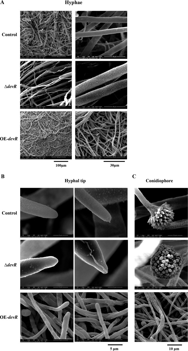
FIG 2.
Electron microscopy. (A) Observation of hyphal morphology. The control, the OE-devR, and the ΔdevR strains were cultivated on CD (dextrin) plates at 30°C for 2 days. (B and C) Further observation of the hyphal tip (B) and conidiophore (C). The marginal regions of the colonies were cut, fixed, and dehydrated. The specimens were examined using an environmental scanning electron microscope. ΔdevR, AodevR-disrupted strain; OE-devR, AodevR-overexpressing strain.

