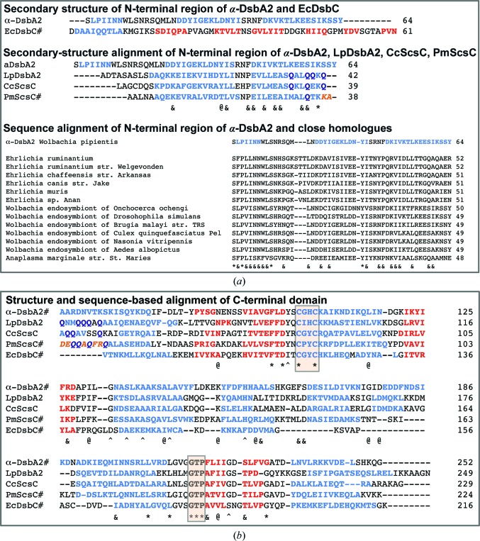
Figure 1.
Comparison of the sequences and structures of bacterial disulfide isomerases. (a) Top panel: alignment of the N-terminal regions of α-DsbA2 (excluding the signal peptide residues 1–15) and EcDsbC. Secondary structure was determined from the structure of EcDsbC (PDB entry 1eej) or was predicted using JPred (Drozdetskiy et al., 2015 ▸) for α-DsbA2. Blue, helices; red, strands. Middle panel: sequence alignment of the N-terminal regions of α-DsbA2, LpDsbA2, CcScsC and PmScsC. Secondary structure for α-DsbA2, LpDsbA2 and CcScsC was predicted by JPred and that for PmScsC was determined from the structure (PDB entry 4xvw). Blue, helices; red, strands; orange, the shape-shifter peptide that adopts different conformations in PmScsC (Furlong et al., 2017 ▸), although predicted to be helical by JPred. Dark blue letters indicate glutamine residues in the shape-shifter region of all four proteins. In this alignment, one residue is conserved across the N-terminal regions of all four proteins and is marked ‘*’, one residue is conserved across α-DsbA2, LpDsbA2 and CcScsC and is marked ‘@’, and nine residues are conserved across LpDsbA2, CcScsC and PmScsC and are marked ‘&’. Bottom panel: sequence alignment of the N-terminal regions of α-DsbA2 and close homologues. The homologues were obtained using BLASTp (Altschul & Koonin, 1998 ▸) and were aligned according to sequence matching to α-DsbA2. In this alignment, fully conserved residues are marked ‘*’ and residues that are conserved in at least seven of the 13 α-DsbA2 homologues are marked ‘&’. (b) Structure- and sequence-based alignment of the C-terminal domains of α-DsbA2, LpDsbA2, CcScsC, PmScsC and EcDsbC. The secondary structures of α-DsbA2, PmScsC and EcDsbC are from their structures; the secondary structures of LpDsbA2 and CcScsC are those predicted by JPred. The CXXC active site and cis-proline loop residues are identified by shaded boxes. Sequence colour key: blue, helices; red, strands; orange italics, shape-shifter peptide; dark blue, glutamine residues in the shape-shifter peptide region. In this composite alignment, the ten residues that are conserved in all five proteins are marked ‘*’, the additional seven residues that are conserved across the four proteins α-DsbA2, LpDsbA2, CcScsC and PmScsC (but not EcDsbC) are marked ‘^’, the ten residues that are conserved across the three proteins α-DsbA2, LpDsbA2 and CcScsC (but not PmScsC) are marked ‘@’, and the seven residues that are conserved across LpDsbA2, CcScsC and PmScsC (but not α-DsbA2) are marked ‘&’. In all alignments, the sequences for which structures are known are marked ‘#’.

