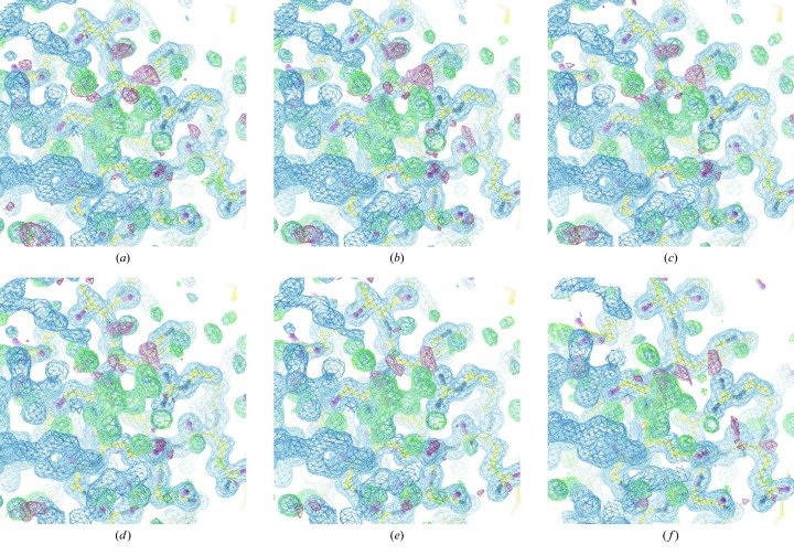
Figure 3.
Difference maps (rendered at 3σ) derived from thaumatin data, showing the tartrate molecule from the crystallization conditions, for data recorded with transmission from  to 64%. Signs of radiation damage are clearly visible in the electron density in the last of these data sets. Of particular interest is the similarity in the maps (b)–(e): by eye there is very little difference in the maps despite the factor of 64 difference in transmission used.

