Figure 4.
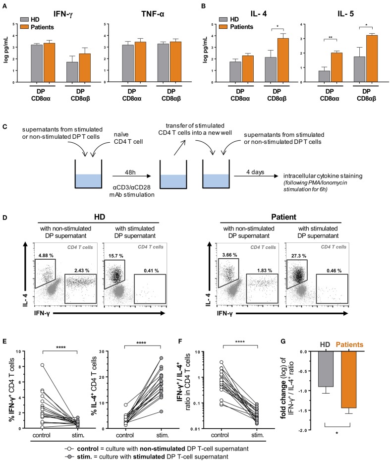
DP T cells favor a Th2 over Th1 polarization of naïve CD4 T cells. DP T-cell clones were generated from ex vivo sorted PBMCs of healthy donors (HD) and urological cancer patients. Clones (3 CD8αα and 4 CD8αβ clones from 3 HD and 6 CD8αα and 4 CD8αβ clones from one patient of each cancer, i.e., bladder, prostate and kidney) were in vitro stimulated (anti-CD3/anti-CD28) for 48 hours, and concentrations (mean ± SEM) of (A) Th1 (IFN-γ, TNF-α) and (B) Th2 (IL-4, IL-5) cytokines were measured in supernatants by Luminex. (C) Experimental procedure. Representative examples (D) and percentages (E) of IL-4 and IFN-γ expression by polarized CD4+ naïve T cells (from 2 HD) upon stimulation in the presence of supernatants from DP T cell clones from HD or patients (described in A,B). (F) Ratio between IFN-γ- and IL-4-expressing CD4+ T cells upon stimulation is shown and (G) variation (fold change) of this ratio (mean ± SEM) compared when using DP T cells from patients and HD. *p ≤ 0.05; **p ≤ 0.01; ****p ≤ 0.0001.

