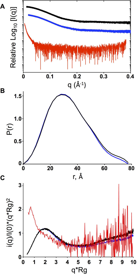
Figure 4.

Small angle X-ray scattering (SAXS) analysis of PMS2 WT, p.Gly207Glu, and p.Leu42_Glu44del proteins. A) Scattering intensity curves for PMS2 WT (black), p.Gly207Glu (blue), and p.Leu42_Glu44del (red). B) Normalized pairwise interatomic distance distribution P[r] function for WT and p.Gly207Glu proteins. C) Kratky analysis indicating the degree of disorder for WT, p.Gly207Glu and p.Leu42_Glu44del.
