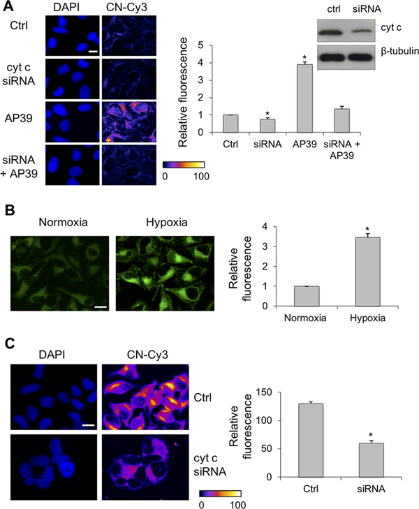
Figure 4.
Cyt C silencing decreases protein persulfidation. (A) Under normoxic conditions, silencing of Cyt C in HeLa cells (confirmed by Western blot analysis) led to a small but measurable decrease in basal persulfidation levels, which was increased by the mitochondrially targeted H2S donor, AP39 (200 nM). The scale bar is 5 μm. (B) Exposure of HeLa cells to hypoxia for 1 h results in increased endogenous H2S levels as detected by MeRho-Az. The scale bar is 10 μm. (C) Under hypoxic conditions, the levels of protein persulfidation are high but significantly reduced by Cyt C silencing. The scale bar is 10 μm. Representative microscopy images are given on the left, and quantitative image analysis is given on the right. The results represent the average fluorescence intensity (F. I.) detected from ≥40 cells ± the standard error of the mean of five independent images (*p < 0.001).

