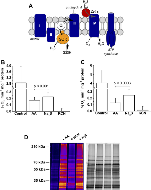
Figure 5.
Sulfide stimulates O2 consumption by mammalian cells and controls persulfidation of mitochondrial proteins. (A) Cartoon showing electrons from H2S oxidation enter the ETC at the level of complex III via the reduced quinone pool. Entry into complex IV via Cyt C can be monitored in the presence of antimycin A, a complex III inhibitor. (B and C) O2 consumption rates of HT29 and HepG2 cells, respectively, before (control) and after treatment with 2 μg mL−1 antimycin A (AA) followed by 20 μM Na2S. Finally, antimycin A-treated cells were exposed to 5 mM KCN. The data are means ± SD of 12 (B) or 6 (C) independent experiments; the p value shows the statistical significance for the difference between antimycin A with and without sulfide-treated cells. (D) Protein persulfidation in purified mitochondria is affected by the ETC. Functional mitochondria were isolated from S. cerevisiae and preincubated with 2.5 μg mL−1 antimycin A or 10 mM KCN for 10 min at 37 °C or treated with 1 μM H2S for 20 min at 37 °C. Persulfidation was monitored by the improved tag swtich method. The first lane represents the basal persulfidation level in untreated functional mitochondria. The total protein load is shown in the gel on the right.

