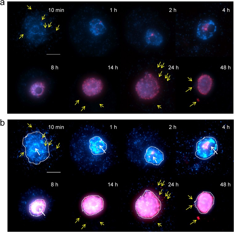
FIG 3.
Time-dependent fluorescence in situ hybridization (FISH) analysis of medusavirus DNA localization in medusavirus-infected A. castellanii cells. Red, medusavirus DNA labeled by Cy-3; blue, both A. castellanii genomic DNA and medusavirus DNA stained by DAPI (4′,6-diamidino-2-phenylindole). Yellow arrows indicate medusavirus DNA signals in the cytoplasm of host cells. The sampling times after virus infection are indicated on the top right corner of each panel. Bar, 10 μm. (a) Original images. (b) Brighter images. Contrast was digitally enhanced. Dashed lines indicate host cell nucleus.

