FIG 5.
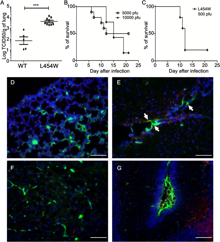
In vivo infection with virus bearing the L454W F. (A) Cotton rats (n = 8) were infected intranasally with MeV IC323-EGFP and MeV IC323-EGFP F L454W F viruses and were euthanized at 4 days postinfection. MeV titration of lung homogenates showed that L454W F bearing virus grew to a significantly higher titer than wt virus in cotton rats (P = 0.001 by the Mann-Whitney U test). The limit of viral detection was 102 median tissue culture infective dose per gram of tissue (TCID50/g). (B) CD150/SLAM suckling mice were infected intranasally with either 5,000 (n = 10) or 10,000 (n = 7) PFU of MeV IC323-EGFP. (C) CD150/SLAM suckling mice were infected intranasally with 500 PFU of MeV IC323-EGFP-F L454W (n = 5). Cryosections of lungs (D) and brain areas (E to G) collected from CD150/SLAM suckling mice (n = 3) coinfected intranasally with 1,000 PFU of both MeV IC323-tdTomato and MeV IC323-EGFP-F L454W (at day 4 postinfection) were stained using anti-GFP (green) and anti-tdTomato (red) antibodies. Nuclei were counterstained with DAPI (blue). (D) Lung section. (E) Brain ventricle area (white arrows indicate the infection in meninges). (F) Cortex parenchyma. (G) Cerebellum. Scale bars, 100 μm.

