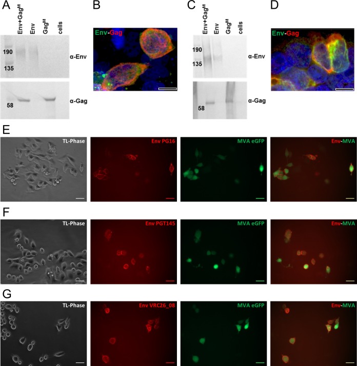
FIG 2.
DNA and rMVA vaccine characterization. Western blots demonstrate in vitro secretion of Env and Gag protein from HEK293 cells after DNA vaccine transfection (A) or MVA vaccine infection (C). “Cells” refers to untransfected and uninfected cells. Membranes were cut in half and the top was probed with anti-Env antibodies and the bottom with anti-Gag antibodies. Confocal images show that both Env (green, Cy3) and Gag (red, Alexa Fluor 647) were expressed in the same cell when DNA vaccines were cotransfected (B) or when infected with rMVA containing both gp150 and GagM (D). Scale bars in confocal images represent 10 µm. (E to G) Live-cell staining of HeLa cells infected with MVA Env, using MAbs PG16 (E), PGT145 (F), and CAP256 VRC26_08 (G), which specifically detect native-like, trimeric Env. HeLa cells infected with rMVA were visualized by their eGFP expression (green). MAbs were detected with anti-human IgG-Cy3 (red). TL-Phase, transmitted light, phase contrast. Scale bars represent 20 µm.

