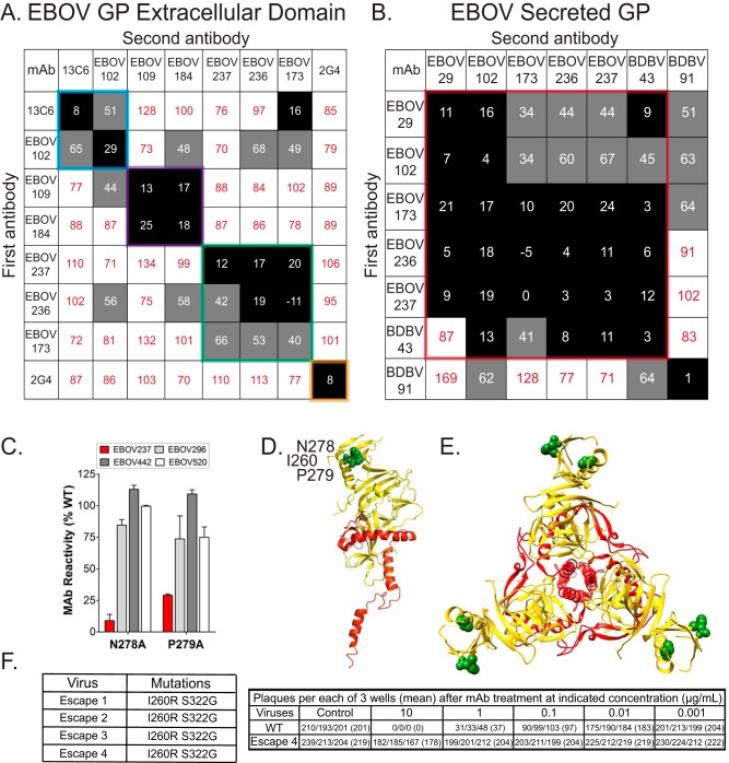
FIG 3.
Epitope mapping of EBOV237 indicates binding to the glycan cap. (A and B) Competition-binding analysis of MAbs to full-length EBOV GP (A) and EBOV sGP (B) are indicated. The numbers indicate residual percent binding of the second antibody in the presence of the first antibody, in comparison to the absence of the first antibody. A reduction in percent binding of the second antibody to <30% due to the presence of the first antibody (black boxes with white numbers) indicates full competition for binding. A reduction in percent binding of the second antibody from 30% to 70% due to the presence of the first antibody (gray boxes with black numbers) indicates intermediate competition. A reduction in percent binding of the second antibody to <30% due to the presence of the first antibody (white boxes with red numbers) indicates a lack of competition. (C to E) EBOV237 was epitope mapped by screening on an EBOV Δmucin GP alanine scan mutation library expressed in HEK-293T cells, with binding by EBOV237 assayed by flow cytometry. This identified N278A and P279A mutants as critical clones that showed specifically reduced binding for EBOV237 Fab (<20% and <30% of binding to WT EBOV GP, respectively; red bar) but a high level of binding to control MAbs EBOV296, EBOV442, or EBOV520 (gray/white bars) (C). Error bars represent the mean and range (half of the maximum minus minimum values) of at least two replicate data points. (D and E) The monomer (D; side view) or trimer (E; top view) form of EBOV Δmucin GP, with GP1 in yellow and GP2 in red (PDB 5JQ3) is shown (31). The critical binding residues for EBOV237 (N278, P279, and I260) to EBOV Δmucin GP are indicated by the green spheres on either structure. S322 is not shown, as this residue corresponds to the mucin-like domain of EBOV GP. (F) Four antibody neutralization escape mutant viruses were isolated, each with the mutations I260R and S322G. A PRNT was done using 10, 1, 0.1, 0.01, or 0.001 μg of EBOV237 incubated with either wild-type (WT) virus or escape mutant 4. The numbers for the assays, performed in triplicate, indicate the PFU per well.

