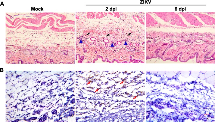
FIG 3.
Histopathological and virological characterization assays in the skin of tree shrews. Skin tissues were collected from ZIKV-infected tree shrews at the indicated time points. (A) Histopathological characterization was performed by hematoxylin and eosin (H&E) staining. The arrows denote areas of hemorrhage, and the triangles denote inflammatory cell infiltration. (B) ZIKV genome RNA ISH was performed with a ZIKV-specific probe. Brown-colored staining indicates positive results (arrows). Bar, 50 μm.

