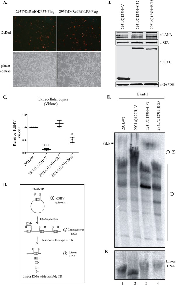
FIG 5.
EBV-BGLF5 only partially complements the abolished DNase activity of the Q129H mutant. (A) HEK293T cells transfected with either the pLVXDsRedORF37-Flag or pLVXDsRedBGLF5-Flag lentiviral construct and imaged by fluorescence microscopy for enhanced DsRed expression at 72 h posttransfection. Red indicates the expression of DsRed, confirming the presence of ORF37 and BGLF5, and the gray images are the corresponding bright-field images. (B) 293L/Q129H cells transduced with empty vector (pLVXDsRed-Flag) or with the indicated lentiviruses and selected with puromycin to generate 293L/Q129H cells stably expressing ORF37-wt (293L/Q129H+C37), BGLF5 (293L/Q129H+BG5), or empty vector (293L/Q129H+V) as a control. Western blot analysis of protein extracts from 48-hpi 293L/Q129H-V, 293L/Q129H+C37, and 293L/Q129H+BG5 cells showed equivalent LANA and RTA expression levels, as well as the respective stably expressed proteins with anti-Flag antibody. The GAPDH immunoblot shows equal loading of the cell lysates. (C) Extracellular KSHV progeny virions assessed from 5-dpi 293L/wt, 293L/Q129H+V, 293L/Q129H+C37, and 293L/Q129H+BG5 cells showed substantial but not complete complementation by the BGLF5 protein of EBV. *, P < 0.1; ***, P < 0.001. Horizontal lines represent the means for triplicate samples. (D) Schematic of the KSHV genome concatemer following BamHI digestion. (E) Southern blot analysis of BamHI-cleaved DNA fragments from 72-hpi 293L/wt, 293L/Q129H+V, 293L/Q129H+C37, and 293L/Q129H+BG5 cells, as analyzed by PFGE and hybridization with a 32P-labeled TR probe. BGLF5 complementation of 293L/Q129H cells only partially restored the abolished DNase activity of ORF37. “1” and “2” represent circular and concatemeric DNAs; “3” corresponds to linear DNA segments. (F) Gardella gel electrophoresis to resolve the linear KSHV genome following BamHI digestion, Southern blotting. and hybridization with a 32P-labeled TR probe.

