
Figure 3.
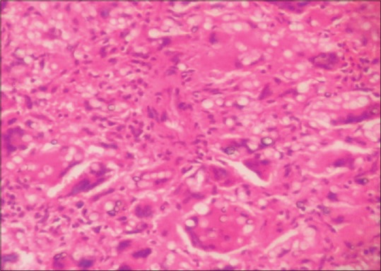
This photomicrograph shows numerous multinucleated giant cells containing cytoplasmic round-to-oval fungal bodies. Furthermore, it observes numerous histiocytes with eccentrically located nuclei with cytoplasmic vacuoles containing oval-shaped fungal bodies of Histoplasma duboisii. Similar bodies were also seen in the giant cells and also extracellularly (H and E, ×300)