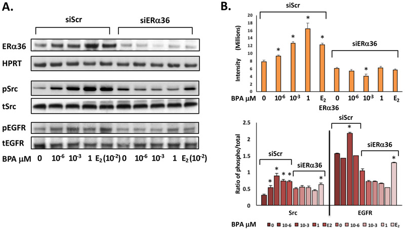
Fig 4. ERα36 mediates the activation of Src, EGFR, and MAPK in ht-UtLM cells exposed to BPA.
(A) Representative western blots and (B) band intensity bar graphs of ERα36 and phosphorylated over total (phospho/total) ratios of intermediary proteins (Src) and the receptor tyrosine kinase (EGFR) in ERα36 knockdown (siERα36) or scrambled RNA (siScr) transfected ht-UtLM cells exposed to BPA for 24h or 10 min. There were significantly (*P<0.05) higher expression levels of ERα36, phospho-EGFR (pEGFR), and phospho-Src (pSrc) in the cells treated with BPA with a functional ERα36 (siScr) compared to controls. However, all elevated protein and phosphorylation levels were diminished when ERα36 was knocked down (siERα36). The western band images shown are representative of three independent experiments and the data were expressed as mean±SE done in three independent experiments.

