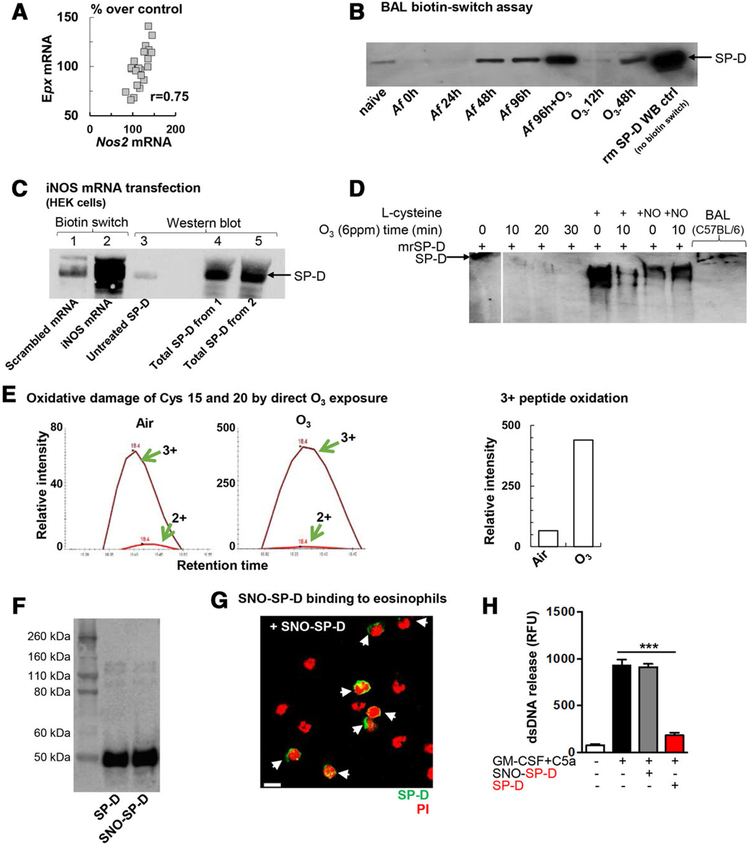
FIGURE 4. S-nitrosylation of the SP-D molecule altered its structure and abolished its inhibitory effects on dsDNA release in a carbohydrate dependent manner.
(A) Correlation between eosinophil peroxidase (Epx) and inducible nitric oxide synthase (Nos2) mRNA in the lung. Data points represent individual Balb/c mice that received the combination of Af sensitization and challenge +O3 exposure (n = 21). (B) Representative blot of n = 3 independent experiments demonstrating S-nitrosylated SP-D in the BAL fluid of allergen and O3 exposed mice by the biotin-switch assay. Mouse recombinant SP-D was added as a Western Blot control without the biotin switch. Naïve: no treatment; O3: ozone exposure (2 ppm for 2h, BAL harvested 12 or 48h later); Af: BAL harvested as indicated; Af+O3: sensitized mice were challenged with Af and received O3 exposure 84 h later. BAL was harvested 96 h after allergen challenge (i.e.,12 h after O3). Rm (recombinant mouse) SP-D: 0.05μg (C): S-nitrosylation of recombinant SP-D with NO obtained by iNOS mRNA translation in HEK cells in vitro. HEK 293 cells were transfected with iNOS mRNA and incubated with rSP-D. After overnight incubation, cell culture supernatants were processed for biotin switch assay. 1, control (scramble transfection)+rSP-D (3 μg)-(biotin switch); 2, iNOS mRNA transfection+ rSP-D (3 μg)-(biotin switch); 3, untreated SP-D (50 ng) –WB ctrl; 4, total SP-D from Sample 1 (WB ctrl, no biotin switch); 5, total SP-D from sample 2 (WB ctrl, no biotin switch). (D) effect of O3 exposure, l-Cys and l-Cys-NO treatment on the structure of recombinant SP-D in vitro. Native gel electrophoresis. (E) Oxidation of peptide SVPNTCTLVMCSPTENGLPGR after in vitro ozone treatment. Recombinant mouse SP-D (3 μg) was de-glycosylated, trypsinized overnight, and then assessed by the TSQ-Vantage mass spectrometer. (F) Immunoblot analysis of denatured recombinant native and oxidized (SNO) SP-D. Results are representative of at least 3 independent experiments. (G) Confocal microscopy. Immunofluorescent staining of bound SNO-SP-D molecules (at 5 μg/mL) on the surface of mature mouse eosinophils isolated from bone marrow of IL5tg mice. Mouse eosinophils were stained with anti-His-Tag Ab. SNO-SP-D binding is indicated by white arrows; scale bar, 10 μm. (H) Quantification of released dsDNA in supernatant of non-activated and activated eosinophils pre-incubated with recombinant SP-D or oxidized SP-D (SNO-SP-D) both at 10 μg/mL. Mean ± SEM of n = 4

