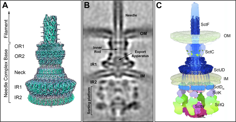
Figure 1.

Salmonella Typhimurium SPI-1 encoded type III secretion system. (A) Surface view of the 3D reconstruction of the single particle cryo-EM map of the needle complex substructure with the atomic structures of the different needle complex components docked. OR1: outer ring 1; OR2: outer ring 2; IR1: inner ring 1; IR2: inner ring 2. (B) Central section of an overall cryo-ET structure of the complete injectisome in situ. Of note is the location of IR2 in the cytosolic side of the bacterial envelope. (C) Molecular model of the organization of the injectisome in situ with available atomic structures fitted into the model (figure adapted from DOI: 10.1016/j.cell.2018.01.034)
