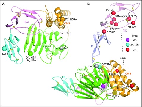
Figure 7.
Important histidines in D assemblies, specific VW type 2A and 2N disease mutations, and FVIII binding model. (A) Conserved histidines in D assemblies that when mutated, diminish or abolish D3 dimerization. Four histidines in D2, 1 in D1, and none in D3 abolish dimerization when mutated to alanine.21 These histidine residues are labeled next to the positions of the homologous residues in D3 which are shown as Cα atom spheres. (B) VW disease 2A and 2N mutations that are likely to be specific: 2A and 2N mutations that represent mutation of Cys or to Cys or are also reported as type 1 or 3 mutations have been excluded. Positions of mutations are shown as Cα atom spheres color-coded as in the key to right. Mutated residues are labeled. The distance from the centers of mutations in TIL′ and C8-3 is shown as a dashed line.

