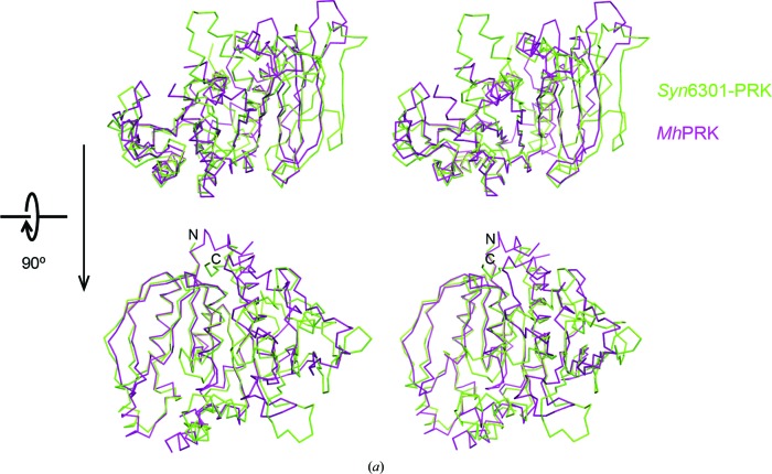
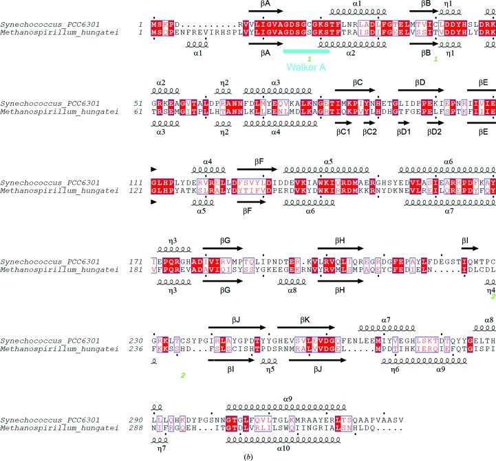
Figure 5.
Comparison of Syn6301-PRK and MhPRK protomers. (a) Stereo representation of superposition of the protomers of Syn6301-PRK and MhPRK. Two perpendicular views are shown. The Cα traces of Syn6301-PRK and MhPRK are shown in green and purple, respectively. The same orientations as in Fig. 4 ▸ are shown. (b) Structure-based sequence alignment of Syn6301-PRK and MhPRK. Secondary-structure elements of the Syn6301-PRK and MhPRK structures are indicated above and below the sequences, respectively. Similar residues are shown in red and identical residues are shown in white on a red background. Green numbers below the sequence indicate the disulfide bonds in redox-blocked Syn6301-PRK. The Walker A motif is indicated in cyan. This figure was prepared using ESPript (Gouet et al., 1999 ▸).


