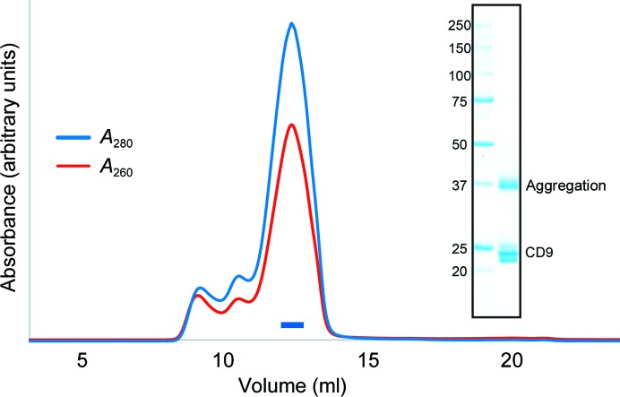
Figure 1.
Protein preparation. Size-exclusion chromatogram of wild-type CD9. The blue and red lines indicate the absorbance at 280 and 260 nm, respectively. The blue bar indicates the fractions that were collected and used for crystallization. The inset shows an SDS–PAGE analysis with Coomassie Brilliant Blue staining. Left lane, molecular-mass markers (labeled in kDa); right lane, wild-type CD9. The multiple bands at around 23 kDa represents heterogeneous palmitoylation of the purified CD9 protein, and a higher molecular-weight band at 37 kDa is owing to molecular aggregation during SDS denaturation.

