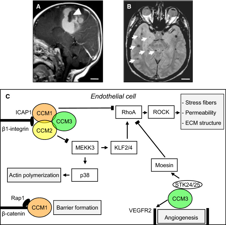
Fig. 1.
Cerebral cavernous malformations (CCMs). a Sagittal magnetic resonance (MR) imaging shows a large cavernoma (arrowhead) with a resultant gross hemorrhage. b Axial MR image of a patient with the genetic form of CCM demonstrates multiple small lesions with a characteristic hemosiderin ring surrounding them (arrows). Scale bars (a, b): 2 cm. Images courtesy of Dr. Murat Gunel (Department of Neurosurgery, Yale School of Medicine and Yale New Haven Hospital). c Summary of select interactors, intracellular pathways, and cellular processes that have been associated with the CCM proteins. The three CCM proteins maintain endothelial function and control angiogenesis. CCM1, CCM2, and CCM3 form a trimeric complex with the scaffolding protein CCM2 acting as a hub. All CCM proteins have an antagonistic effect on the RhoA/ROCK signaling pathway mediated by distinct interactors (arrow-headed lines: activation; bar-headed lines: inhibition). RhoA/ROCK pathway activation in the absence of CCM proteins results in stress fiber formation, increased vascular permeability (possibly explaining why lesions in patients tend to leak causing microhemorrhages) of CCM, and alters the composition of the extracellular matrix (ECM). CCM1 interaction with ICAP1 inhibits β1-Integrin activation and supports Rap1-mediated stabilization of endothelial junctions, thereby contribution to barrier formation. CCM2 interaction with MEKK3 results in decreased p38 activity to modulate actin polymerization. CCM3 stabilizes VEGFR2 signaling to regulate angiogenesis and negatively regulates Rho via its direct interactors STK24/25, which activate moesin. CCM: cerebral cavernous malformation. ECM: extracellular matrix. ICAP1, integrin cytoplasmic domain-associated protein 1; KLF2/4, Krüppel-like factor 2/4; MEKK3, mitogen-activated protein kinase kinase kinase 3; Rap1, Ras-associated protein 1; RhoA, Ras homolog gene family, member A; ROCK, Rho-associated coiled-coil kinase; VEGFR2, vascular endothelial growth factor receptor 2

