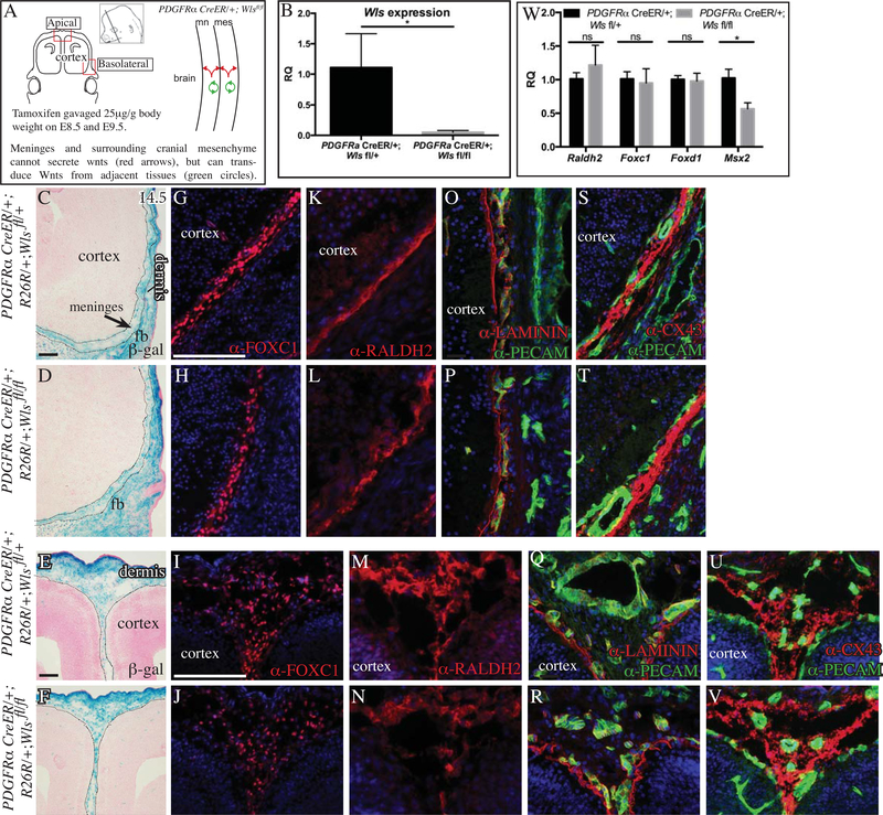
Fig.2. Conditional deletion of Wntless (Wls) in cranial mesenchyme.
Schematic illustration of the coronal plane, tamoxifen induction regimen, lateral view of the embryonic head in the region of interest, and simplified schematic of genetic model (A). Relative quantity (RQ) of mRNA in E14.5 cranial mesenchyme of control and conditional Wls mutant cranial mesenchyme (n=4) (B, W). β-galactosidase staining with eosin counterstaining and black dotted lines outline meningeal mesenchyme (C-F). Indirect immunofluorescence for markers of meninges (G-N), basement membrane LAMININ (red, O-R), CD31/PECAM+ endothelial cells (green, O-V), Connexin43 in gap junctions (red, S-V) with DAPI-stained (blue) nuclei (G-V). Scale bars represent 100μM. * indicates statistical significance with p value between 0.01 and 0.05 (B, W). White and black dotted line demarcate the meningeal mesenchyme from adjacent cranial mesenchyme and cortex.

