. 2019 Apr 5;8:e42434. doi: 10.7554/eLife.42434

Figure 1. Mosaic membrane GFP labeling of nascent mesoderm allows following individual cell migration through embryo live imaging.

(a) Posterior view. Top: 3D scheme with mesoderm layer in green and the rest of the embryo in grey. The dashed line separates embryonic and extra-embryonic regions. Middle: Z-projection of two-photon stack. Bottom: optical slice highlighting the primitive streak. (b) Lateral view, anterior to the left. Top: 2D scheme. Middle: Z-projection of two-photon stack showing cells progression from posterior to anterior. Bottom: sagittal optical slice. (c) Anterior view. Top: 3D scheme. Middle: Z-projection of two-photon stack with most anterior cells reaching the midline. Bottom: optical slice zoomed on filopodia extending towards the midline. All embryos were dissected at E7.25 and are at Late Streak/Early Bud stage. VE: Visceral Endoderm; mG: membrane GFP, in green; mT: membrane dtTomato, in grey; EB: Early Bud; LS: Late Streak; 0B: Zero Bud. (Scale bars: 50 μm).

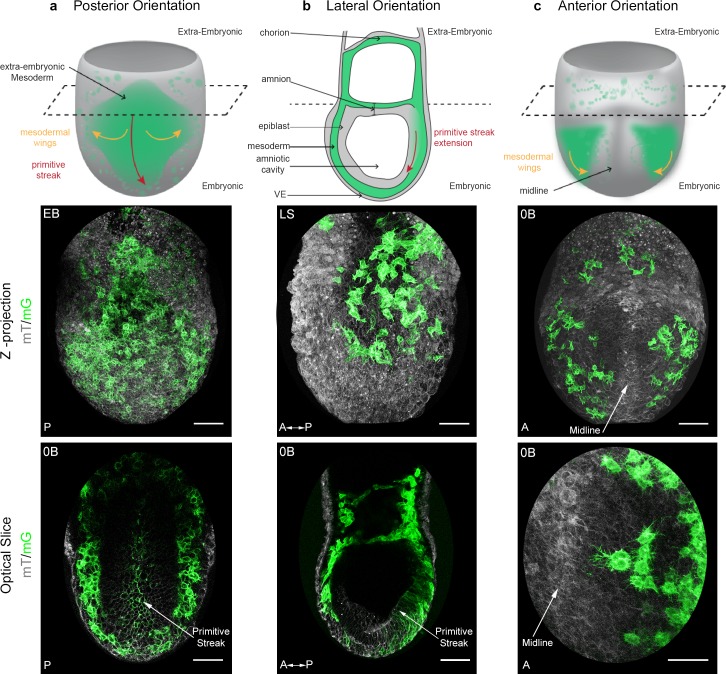
Figure 1.

Figure 1—figure supplement 1. Live imaging of Brachyury (T)-Cre; mTmG embryos.

Figure 1—figure supplement 1.

(a) Sketches showing mouse embryo anatomical landmarks used for staging between Early Streak and Early Bud stages. (b) Quantification of embryonic and extra-embryonic mesoderm cell division events (mean ± SEM, n=4 embryos, p=0.0286, Figure 1—figure supplement 1—source data 1.). Cells were observed over an average of 360 min. Cells migrating close to the embryonic/extra-embryonic interface were not taken into consideration. (c) Ex vivo growth of an embryo dissected at E6.75 (Early Streak stage, left panel), after imaging for 12 hr using confocal imaging plus 12 hr of subsequent culture (Zero Bud stage, right panel). (d) Ex vivo growth of an embryo dissected at E7.25 (Late Streak stage, left panel), after 8 hr two-photon imaging (Early Bud stage, right panel). (e) Transverse section at primitive streak level of a E7.5 (Late Streak stage) T-Cre; mTmG embryo stained for F-actin (Phalloidin, red) and nuclei (DAPI, blue) (scale bar: 50 μm). (f) Quantification of mosaicism as the ratio of GFP + cells/all cells in the mesoderm layer (identified anatomically between the epiblast and the visceral endoderm) in anterior, posterior and lateral quadrants at different stages of gastrulation: E7 (Mid Streak stage) and E7.5 (Early Bud stage) (mean ± SEM, n=5 embryos for each stage). P values were calculated using the Mann–Whitney–Wilcoxon. A: Anterior, P: Posterior, Pr: Proximal, Di: Distal, mGFP: membrane GFP, in green.
Figure 1—figure supplement 1—source data 1. Division events in mesoderm cells.
DOI: 10.7554/eLife.42434.005