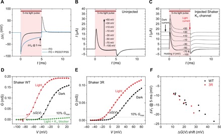
Fig. 4. Photostimulation effects on KVShaker-type channels in oocytes.

(A) Voltage measurements on uninjected oocytes using a 5-ms light pulse in the case where the bare ITO is the back electrode and the ITO is modified with PEDOT:PSS. Because of the higher capacitance given by PEDOT:PSS, the photocapacitive charging currents are larger and are exhibited over longer time, thus applying higher transductive extracellular potentials. Maximizing VT in a time scale of ≥2 ms is critical for observing effective stimulation of KV channels. We use VT@5 ms here as a convenient reference. (B) Voltage-clamp measurements at various voltage steps in uninjected oocytes, showing the voltage-independent capacitive response of the cell to the light pulse. (C) Voltage-clamp measurements of an oocyte transfected with the Shaker KV channel, demonstrating the photoinduced increase in outward K+ current. (D) G(V) characteristic for WT Shaker KV channels, for dark conditions (black trace) and during the light pulse (red trace). The green trace is for measurement after addition of the KV channel blocker 4-AP. (E) G(V) characteristics comparing dark conditions versus light pulses for 3R Shaker mutant channels. The onsets are shifted relative to WT. However, the photoinduced shift behavior is the same. (F) Correlation between the observed voltage shift in the light versus dark G(V) and the transient voltage at the end of a 5-ms light pulse measured in the same cell. The higher transient voltage VT corresponds to greater observed shifts in the G(V) characteristics.
