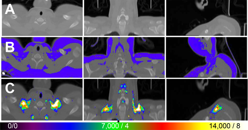
Figure 6.
FDG uptake in PET/CT of a healthy subject at cool temperature. CT scan shows radiodensity of neck and upper chest to just below clavicle (A). Template (blue) indicates all fat superimposed on CT (B). Template was based on radiodensity difference of fat from non-fat tissues (e.g., muscle, lung). FDG uptake in PET/CT scan of control subject resting for 2.5 h in cold (62°F or 16.8°C) (C). Color scale indicates greater uptake with hotter colors. The threshold was set at 3,500 counts (i.e., blue, normalized to whole brain uptake of 20,000) or SUV = 2 based on body weight. Maximum or greater uptake indicated by white color indicating 14,000 counts or SUV 8.

