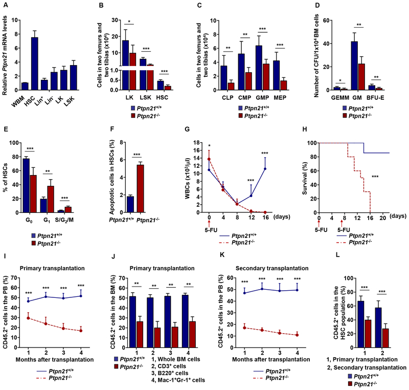
Figure 1. Knock-out of Ptpn21 Results in HSC Defects and Impaired Hematopoiesis.
(A) Lin+, Lin−, LK, and LSK cells and HSCs were sorted from the BM harvested from healthy C57BL/6 mice (n = 5). Total RNA was extracted from these purified cells and whole BM (WBM) cells. Ptpn21 mRNA levels were determined by qRT-PCR. (B and C) BM cells freshly harvested from 6–8 week old Ptpn21−/− and Ptpn21+/+ mice were assayed by multiparameter FACS analyses to determine the numbers of LK cells, LSK cells, and HSCs (n = 6 mice per genotype) (B), and CLPs, CMPs, GMPs, and MEPs (n = 7 mice per genotype) (C). (D) BM cells harvested from Ptpn21−/− and Ptpn21+/+ mice (n = 5 per genotype) were assessed by CFU assays. Hematopoietic colonies (CFU-GEMM, CFUGM, and BFU-E) were enumerated. (E and F) BM cells harvested from Ptpn21−/− and Ptpn21+/+ mice (n = 6 per genotype) were assayed by FACS for the cell cycle status and apoptosis in HSCs. Percentages of HSCs in G0, G1, and S/G2/M phases (E) and apoptotic cells (Annexin V+) in HSCs (F) were quantified. (G) Six to eight-week-old Ptpn21−/− and Ptpn21+/+ mice (n = 10 per genotype) were administrated 5-Fluorouracil (5-FU) [250 mg/kg body weight, intraperitoneal (i.p.)]. White blood cell (WBC) counts in the PB were monitored. (H) Ptpn21−/− (n = 10) and Ptpn21+/+ (n = 11) mice were administrated two doses of 5-FU (150 mg/kg body weight, i.p.) at one week interval. Animal survival rates were determined. (I) BM cells (test cells) harvested from Ptpn21−/− or Ptpn21+/+ mice (CD45.2+) were mixed with BoyJ (CD45.1+) BM cells (1:1 ratio) and transplanted into lethally irradiated BoyJ recipients (n = 10 per genotype). Test cell reconstitution in the whole cell population of the PB at the indicated time points was determined by FACS analyses. (J) Test cell reconstitution in the whole cell population and in each lineage in the BM was determined 4 months after primary transplantation (n= 8 mice per genotype). (K) BM cells harvested from recipients 4 months after primary transplantation were transplanted into lethally irradiated secondary BoyJ recipient mice (n = 9 per genotype). Test cell reconstitution in the PB of secondary recipients was determined at the indicated time points as above. (L) Test cell contributions to the BM HSCs were determined 4 months after primary and secondary transplantation by multiparameter FACS analyses (n = 9 mice per genotype). See also Figures S1, S2, and S3.

