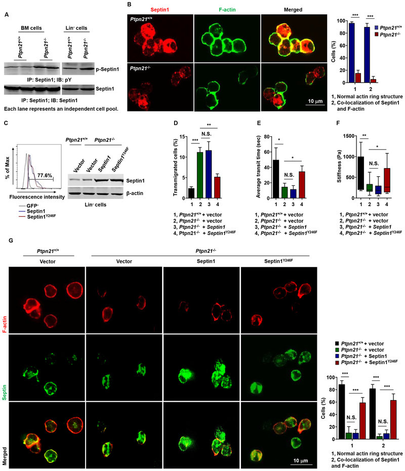
Figure 7. Ptpn21 Maintains Cell Mechanics by Dephosphorylation of Septin1 (Tyr246).
(A) BM cells and Lin− cells freshly isolated from Ptpn21−/− and Ptpn21+/+ mice were lysed and examined by immunoprecipitation followed by immunoblotting analyses. Representative results from at least 3 independent experiments are shown. (B) HSCs isolated from Ptpn21+/+ and Ptpn21−/− mice (n = 4 per genotype) were processed for immunofluorescence staining for Septin1 and F-actin. Representative confocal images and quantification of the percentages of cells with normal F-actin structure and F-actin/Septin1 co-localization (90–120 cells examined per mouse sample) are shown. (C-G) Lin− cells isolated from Ptpn21+/+ and Ptpn21−/− mice (n = 3–4 per genotype) were infected with lentiviruses expressing WT Septin1 or Septin1Y246F. Forty-eight hours later, infected cells were assessed for transduction efficiencies by FACS analyses of GFP and for expression levels of Spetin1 by immunoblotting (C). Lentiviruses-infected Lin− cells were also assessed by transwell (3 μm pores) migration assays in the presence of SDF-1 (D), microfluidic assays (E), AFM measurements of cell stiffness (F), and immunofluorescence staining for Septin1 and F-actin. Representative confocal images and quantification of the percentages of cells with normal F-actin structure and F-actin/Septin1 co-localization (90–120 cells examined per mouse sample) are shown (G). See also Figures S6 and S7.

