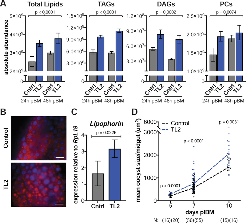
Figure 7: Silencing the TAG lipase TL2 induces faster parasite growth.
(A) Total lipids, TAGs, DAGs, and PCs are higher in dsTL2 midguts (TL2) at 24 h and 48 h pBM compared to dsGFP controls (Cntrl) (means ± SEM, Least Squares model). (B) Midguts of dsTL2 females stained with the neutral lipid dye LD540 (red) show an accumulation of lipid droplets at 48 h pBM (scale bar = 20 μm). Blue = DAPI. (C) Lipophorin expression is elevated in dsTL2 fat bodies at 24 h pBM (means ± SEM, Least Squares model). (D) dsTL2 females develop larger oocysts from 5–10 d pIBM (Mann-Whitney). N = sample size with dsGFP listed first. See Figures S4–S5.

