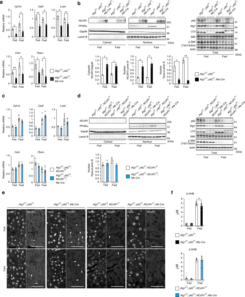
Fig. 5.
NCoR1-dependent PPARα-inactivation in autophagy-deficient livers. a, c Gene expression of enzymes related to lipid oxidation in Atg7 p62 double- (a) and Atg7 p62 NCoR1 triple-knockout livers (c). Total RNAs were prepared from livers of Atg7f/f;p62f/f (fed: n = 3; fasted: n = 5), Atg7f/f;p62f/f;Alb-Cre (fed: n = 3; fasted: n = 4), Atg7f/f;p62f/f;NCoR1f/f (fed: n = 4; fasted: n = 4), and Atg7f/f;p62f/f;NCoR1f/f;Alb-Cre (fed: n = 4; fasted: n = 4) mice aged 5 weeks under both fed and fasting conditions. Values were normalized against the amount of mRNA in the livers of fasting Atg7f/f;p62f/f or Atg7f/f;p62f/f;NCoR1f/f mice. The experiments were performed three times. b, d NCoR1 level in Atg7 p62 double- (b) and Atg7 p62 NCoR1 triple-knockout livers (d). Total homogenate, as well as nuclear and cytoplasmic fractions, were prepared from livers of mice of the indicated genotypes and subjected to immunoblotting with the indicated antibodies. Bar graphs indicate the quantitative densitometric analyses of indicated cytoplasmic and nuclear proteins relative to Gapdh and Lamin B, respectively. e Immunohistofluorescence microscopy. Liver sections of Atg7f/f;p62f/f, Atg7f/f;p62f/f;Alb-Cre, Atg7f/f;p62f/f;NCoR1f/f, and Atg7f/f;p62f/f;NCoR1f/f;Alb-Cre mice aged 5 weeks were immunostained with anti-NCoR1 antibody. Arrowheads indicate intranuclear signals for NCoR1. Bars: 50 μm. f Blood β-OHB in mice described in a. Data are means ± s.e.m. *P < 0.05, **P < 0.01, and ***P < 0.001 as determined by Welch’s t-test

