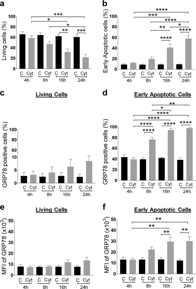
Fig. 2. Surface translocation of glucose-regulated protein 78 (GRP78) in beta cells is an early phenomenon, taking place in the early apoptotic cell population.
MIN6 cells exposed to cytokines (human interleukin-1β (50 U/ml), mouse interferon-γ (250 U/ml), and mouse tumor necrosis factor-α (1000 U/ml)) for the indicated time points (4, 8, 16, and 24 h) were stained with the late-apoptosis/necrosis marker DRAQ7-AF700, early apoptosis marker Annexin V-APC (see Results for the interpretation of these combined dyes to discriminate between living, early, and late apoptotic cells), and anti-GRP78-FITC, followed by flow cytometry. a, b Percentage of living (a) and early apoptotic (b) cell population in control (C) and cytokine (Cyt) exposed MIN6 cells. c, d Percentage of sGRP78-positive cells in the living (c) and early apoptotic (d) subpopulation. e, f Geometric mean fluorescence intensity (MFI) for sGRP78 in the living (e) and early apoptotic (f) subpopulation. Data are presented as mean ± SEM (n = 4) and statistically analyzed by a two-way analysis of variance followed by Sidak posthoc test for multiple group comparisons. *P < 0.05, **P < 0.01, ***P < 0.001, and ****P < 0.0001

