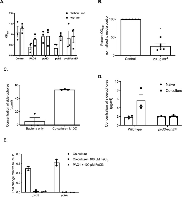
Figure 5.
P. aeruginosa-imposed iron restriction is largely mediated via pyoverdine production. (A) R. microsporus was grown in bacterial supernatants from wild type P. aeruginosa, strains defective in siderophore biosynthesis, or standard LB mixed 25:75 with SAB, with and without iron. R. microsporus spores were exposed to these mixtures for 24 h and fungal growth was determined via absorbance (OD600, n = 3). (B) R. microsporus spores were incubated in SAB with 20 μg ml−1 of exogenous pyoverdine at 37 °C for 24 h. Fungal growth was measured through absorbance (OD600) and normalised to media control (n = 6). (C) P. aeruginosa was exposed to R. microsporus at an MOI of 1:100 (R. microsporus:P. aeruginosa) for 24 h (37 °C). Siderophore production was measured by using the Siderotec Assay (EmerginBio) (n = 3, Mann-Whitney U test). (D) P. aeruginosa strains defective in siderophore biosynthesis were exposed to R. microsporus at an MOI of 1:100 (R. microsporus:P. aeruginosa) for 24 h (37 °C). Siderophore production was measured by using the Siderotec Assay (EmerginBio) (n = 3, Mann-Whitney U test). (E) P. aeruginosa was exposed to R. microsporus at an MOI of 1:100 (R. microsporus:P. aeruginosa) for 7 h, snap frozen and total RNA extracted. The expression levels of PvdS and PchA were quantified by qRT-PCR relative to RpoD and normalised to PAO1 grown in isolation.

