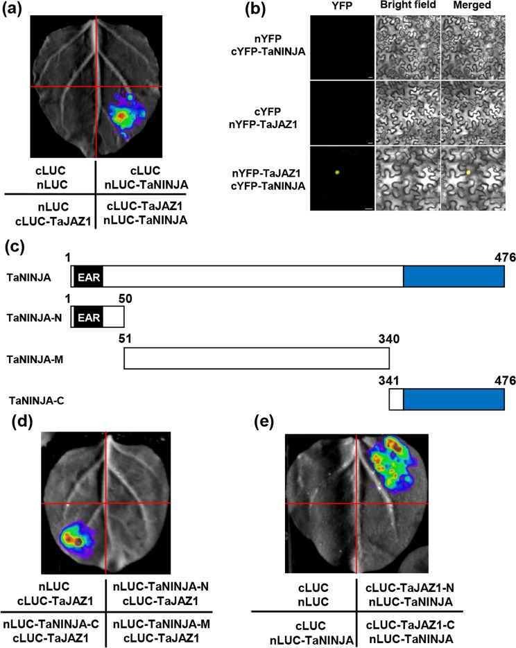
Figure 10.
TaJAZ1 physically interacts with TaNINJA. (a) LCI assay showing the interaction between TaJAZ1 and TaNINJA. EV were used as negative controls. (b) BiFC assay illustrating the TaJAZ1-TaNINJA interaction in the nucleus of N. benthamiana leaf cells. TaJAZ1 and TaNINJA were separately fused with the N- and C-terminal part of YFP (nYFP and cYFP). YFP fluorescence was detected 48 h after infiltration. Bars = 20 µm. (c) Schematic representations of the domain structures of TaNINJA and truncated versions of TaNINJA proteins. (d) LCI assay illustrating that C-terminus of TaNINJA mediates its interaction with TaJAZ1. The nLUC and cLUC EVs were used as negative controls. (e) Mapping of the interaction domain of TaJAZ1 with TaNINJA. The signals of the interaction were collected 48 h after infiltration. Eight N. benthamiana leaves were analyzed in each experiment and similar results were observed.

