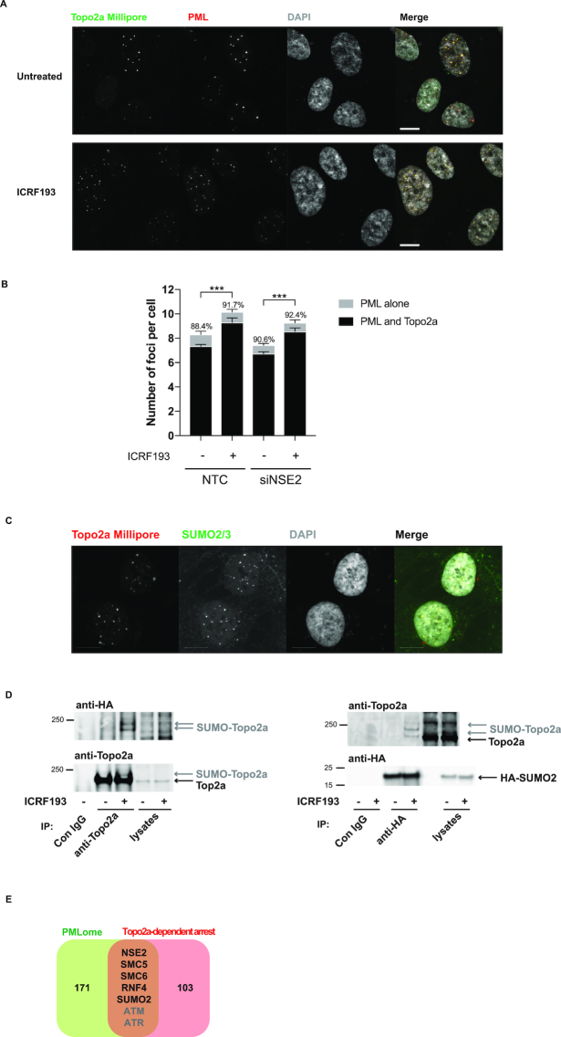
Figure 3.
Topo2a colocalizes with PML bodies and SUMO2/3. (A) IF staining of RPE1 cells for Topo2a and PML after 18 h treatment with 3 μM ICRF193 as indicated. (B) MATLAB-aided quantification, as described in the IF image analysis section of the Materials and Methods, of colocalization between Topo2a (Millipore) and PML (abcam) in the presence and absence of NSE2. RPE1 cells were transfected with non-targeting control (NTC) or siNSE2 for 48 h and subsequently treated with 3 μM ICRF193 for 18 h where indicated. The percentages show the proportion of PML foci that were positive for Topo2a. Data represents mean ± S.E.M. for n = 3. (C) IF staining of RPE1 cells for SUMO2/3 and Topo2a. Scale bar = 10 μm. (D) RPE1 cells were transfected with HA-SUMO2, 24 h later treated with 3 μM ICRF193 for 18 h as indicated before lysates were subjected to either anti-Topo2a (left) or anti-HA (right) immunoprecipitation. Subsequent WB were performed for HA-SUMO and Topo2a. (E) Venn diagram showing overlap between the ICRF193-selective hits and a recently identified PMLome (31). ATM and ATR are redundant players in the Topo2a-dependent G2 arrest.

