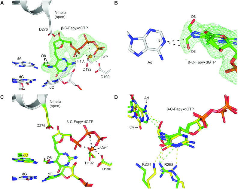
Figure 4.
(A) Pre-catalytic ternary-open complex of wild-type pol β (gray) with β-C-Fapy•dGTP (green sticks) bound across from dA; Ca2+ ions are shown in orange. A polder map (green mesh) contoured at 3.0σ is shown for the incoming β-C-Fapy•dGTP. (B) An alternate viewpoint demonstrating the two formamide conformations of β-C-Fapy•dGTP (green sticks) opposite adenine (gray sticks) with the polder map contoured to 3.0σ (green mesh). (C) An overlay between the pre-catalytic ternary-open structures of β-C-Fapy•dGTP across from dA (green) or dC (yellow). Key residues are shown as gray sticks and the N-helix in gray cartoon. (D) An overlay between the pre-catalytic ternary-open structures of β-C-Fapy•dGTP across from dA (green) or dC (yellow) highlighting altered amino acids in stick format with the N-helix subdomain shown as gray cartoon and potential hydrogen bonds as dashed lines.

