Figure 2.

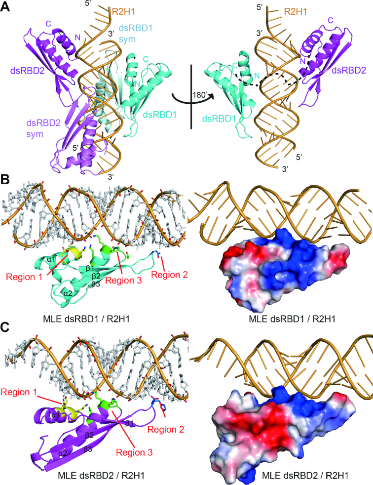
Structural overview of MLE dsRBD1+2 in complex with R2H1. (A) Cartoon representation of the MLE dsRBD1+2–R2H1 complex in two orientations related by a 180° rotation around a vertical axis. Left panel: the asymmetric unit consists of the 55mer R2H1 (bright orange), dsRBD1 (cyan) and dsRBD2 (magenta). Symmetry equivalent domains are shown in pale cyan and violet, respectively. Right panel: a view of the complex with symmetry equivalent protein domains removed for clarity. The linker between the domains is represented as a black dotted line. (B) Overall structure of MLE dsRBD1 in complex with R2H1. Left panel: cartoon view of the structure of MLE dsRBD1 in complex with R2H1. The critical residues belonging to regions 1, 2 and 3 required for dsRNA recognition are shown in stick mode and are colored in yellow, blue and green, respectively. Right panel: the electrostatic potential of the MLE dsRBD1–R2H1 complex is shown, in which positively charged, negatively charged and neutral areas are represented in blue, red and white, respectively. (C) Overall structure of MLE dsRBD2 in complex with R2H1. Left panel: cartoon view of the structure of MLE dsRBD2 in complex with R2H1. The critical residues belonging to regions 1, 2 and 3 required for dsRNA recognition are shown in stick mode and are colored in yellow, blue and green, respectively. Right panel: the electrostatic potential of the surface of the MLE dsRBD2–R2H1 complex, in which positively charged, negatively charged and neutral areas are represented in blue, red and white, respectively.
